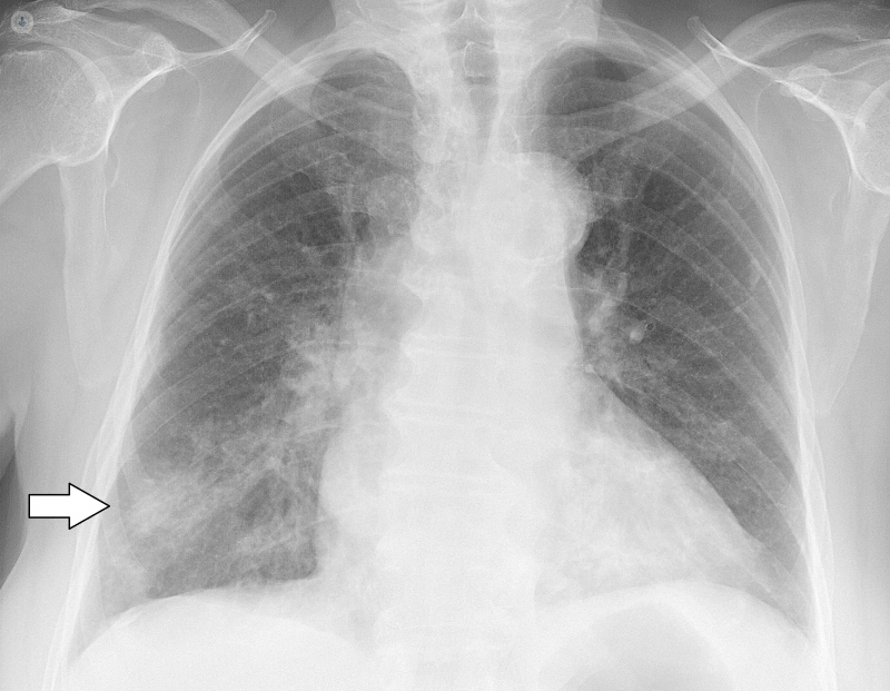
Chest X Ray Normal Means. The standard chest x rays consists of a pa and lateral chest x ray. The dark background on the chest x rays represents air filled lungs.

The image on the right shows a mass in the right lung. On the bottom the chest cavity is bordered by the diaphragm under which is the abdominal cavity. The chest x ray on the left is normal.
On the top portion of the chest are the neck and the collar bones clavicles.
The normal lateral chest x ray view is obtained with the left chest against the cassette. The chest x ray on the left is normal. In a normal chest x ray the chest cavity is outlined on each side by the white bony structures that represent the ribs of the chest wall. If the x ray is a true lateral the right ribs are larger due to magnification and usually projected posteriorly to the left ribs figure 3.